

Understanding, Detecting, and Treating the Deadliest Form of Skin Cancer
Expert diagnosis and a coordinated treatment approach from board-certified dermatologist Dr. Ellen Turner and her care team in Dallas and Irving.
UNDERSTANDING MELANOMA
What Is Melanoma and Why Early Detection Is Everything
Melanoma is the most serious and potentially deadly form of skin cancer. While it is far less common than basal cell or squamous cell carcinoma, melanoma behaves differently, it can spread outward across the surface of the skin, invade downward into deeper tissue layers, and metastasize to other organs, even in otherwise healthy individuals. At the Dermatology Office of Dr. Ellen Turner in Dallas and Irving, we help patients understand their personal risk, catch suspicious lesions early, and coordinate the right treatment team when melanoma is diagnosed.
Melanoma can affect people of any age, race, or ethnic background. No one is exempt. Unlike basal cell and squamous cell carcinoma, melanoma is not caused solely by cumulative UV exposure. While sun protection remains important for every patient, the leading risk factor for melanoma is genetics. A family history of melanoma, or of pancreatic cancer, is a significant signal that a patient should be receiving regular full body skin examinations from a dermatologist.
Melanoma develops when melanocytes, the pigment producing cells that protect skin from UV damage, begin to grow abnormally. For roughly every 15 skin cells, there is one melanocyte, responsible for the pigment that gives skin its color and for shielding nearby cell DNA much like a retractable awning. A large cluster of melanocytes in one area is what we know as a mole, or nevus.
It is estimated that 234,680 cases of melanoma will be diagnosed in the United States in 2026, and roughly 8,510 people will die from the disease this year. Yet when detected and treated before it spreads, the five-year survival rate for melanoma exceeds 99 percent, among the highest of any cancer.

Melanoma at a Glance
- ✓The deadliest form of skin cancer, though far less common than BCC or SCC
- ✓Genetics, not just sun exposure, is the leading risk factor
- ✓Can spread to lymph nodes and distant organs if left untreated
- ✓Detected early, the five-year survival rate exceeds 99 percent
99%+
5-year survival when caught early
234,680
Estimated new U.S. cases in 2026
WHEN TO SEE A DERMATOLOGIST
Know the ABCDEs of Melanoma
Most melanomas are first noticed by patients themselves, not their doctors. Knowing your own skin and checking it regularly is one of the most powerful things you can do.

A
Asymmetry
If a lesion were split down the middle, would both halves mirror each other? If not, that is a warning sign.
B
Border Irregularity
Ragged, notched, or blurred edges, rather than a smooth, well-defined border.
C
Color Variation
Multiple shades of tan, brown, black, or blue-gray within one lesion.
D
Diameter
Often larger than a pencil eraser, about 6mm. Any growing mole deserves evaluation.
E
Evolving
Any mole that is new or changing in size, shape, color, or texture should be evaluated.
REAL PATIENT EXAMPLES

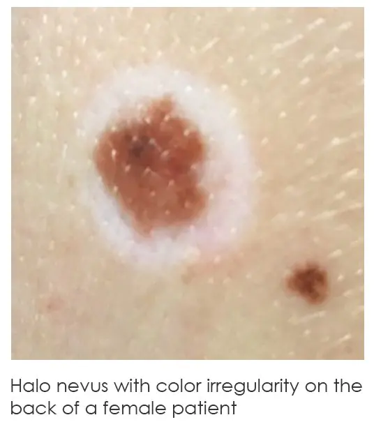
If you notice any of these signs, contact our Dallas or Irving office for an evaluation. Nearly all cutaneous melanomas are first noticed by patients or the people who love them.
SUBTYPES
The Different Forms of Melanoma
Melanoma is not one disease. Dermatologists and dermatopathologists recognize several distinct subtypes, each with its own appearance, growth pattern, and typical location on the body.
MOST COMMON, ~70%
Superficial Spreading Melanoma
Grows outward across the top layer of skin before eventually growing deeper. Often arises from an existing mole and can appear almost anywhere on the body.
MOST AGGRESSIVE
Nodular Melanoma
The second most common subtype, growing quickly and vertically from the start. Typically presents as a firm bump or node, often black or blue-black.
SLOW-GROWING
Lentigo Maligna Melanoma
Develops slowly over years or decades on chronically sun-exposed skin such as the face, scalp, and neck, most common in older adults.
NOT UV-LINKED
Acral Lentiginous Melanoma
Develops on the palms, soles, or under the nails. The most common form of melanoma in people with darker skin tones.
RARE VARIANTS
Amelanotic & Other Subtypes
Amelanotic melanoma lacks visible pigment, often presenting as pink or red. Other rare subtypes include desmoplastic, spitzoid, and nevoid melanoma.
WHY MELANOMA DEVELOPS
Understanding Your Risk
Unlike basal cell and squamous cell carcinoma, melanoma is not driven primarily by cumulative sun exposure. Genetics plays the leading role.
Family History & Genetics
The single greatest risk factor. A family history of melanoma, or of pancreatic cancer, can indicate an inherited CDKN2A gene mutation.
Many or Atypical Moles
Having more than 100 moles, or several atypical (dysplastic) moles, significantly raises lifetime risk.
Fair Skin & Sun Sensitivity
Light skin, light eyes, red or blond hair, and a tendency to burn are all associated with higher risk.
UV Exposure & Sunburn History
Five or more blistering sunburns in a lifetime doubles melanoma risk.
Personal History of Skin Cancer
A prior melanoma, or a prior non-melanoma skin cancer, raises the risk of developing melanoma.
Weakened Immune System
Organ transplant recipients and other immunosuppressed patients face increased melanoma risk.
THE DIAGNOSTIC PROCESS
How a Suspicious Mole Becomes a Confirmed Diagnosis
The gold standard for diagnosing melanoma is a same-day skin biopsy. Here is what to expect at your Dallas or Irving appointment.
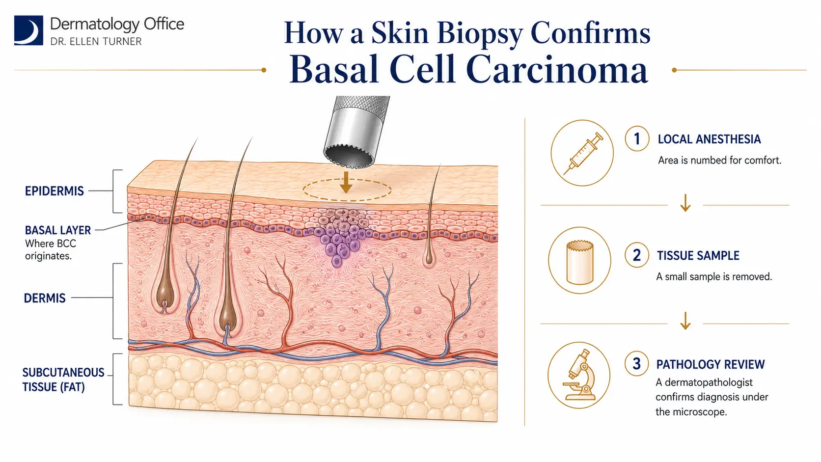
Step 1 – Full-Body Skin Examination
▾
- ✓Dr. Turner performs a thorough visual examination of the lesion in question and any other suspicious moles
- ✓Your personal and family history, including any family history of melanoma or pancreatic cancer, is reviewed carefully
- ✓The ABCDE criteria and any recent changes you have noticed are discussed
- ✓If a biopsy is recommended, the reasons are fully explained before proceeding
Step 2 – Same-Day Skin Biopsy
▾
- ✓The area is cleansed and numbed with a local anesthetic, such as lidocaine, which takes effect within seconds
- ✓A surgical blade is used to remove the tissue sample, typically an excisional or punch biopsy
- ✓The specimen is sent to a fellowship-trained dermatopathologist for microscopic evaluation
- ✓Results are typically available in less than a week, and Dr. Turner personally reviews every report
Step 3 – Staging & Referral
▾
- ✓The dermatopathologist determines the tumor’s depth (Breslow thickness) and whether ulceration is present
- ✓Depending on depth, Dr. Turner may refer you to a surgical oncologist for wide excision and, if indicated, a sentinel lymph node biopsy
- ✓More advanced cases may also involve a medical oncologist as part of a coordinated, team-based treatment approach
- ✓Dr. Turner remains an integral partner in your care throughout treatment and beyond
Step 4 – Ongoing Surveillance
▾
- ✓Full-body skin examinations increase in frequency during the first year after diagnosis, typically every three months
- ✓After the first year, exams continue twice yearly for life
- ✓The frequency of dermatologic skin exams never decreases over a patient’s lifetime after a melanoma diagnosis
- ✓This ongoing surveillance is key to catching any new or recurrent lesions as early as possible
Medical Dermatology
Contact us online about setting up an appointment or call (214) 373-7546. Find out more about what to do about melanoma in the Dallas area today.
TREATMENT APPROACH
A Coordinated, Team-Based Approach to Treatment
Unlike basal cell and squamous cell carcinoma, melanoma treatment is rarely handled by a dermatologist alone. Because melanoma can spread beyond the skin, treatment often involves a team of specialists working together.
Wide Local Excision
The standard surgical treatment for melanoma. The tumor is removed along with a margin of surrounding healthy tissue, with margin size determined by the tumor’s thickness. Typically performed by a surgical oncologist.
Sentinel Lymph Node Biopsy
For melanomas of sufficient thickness, the first lymph node the tumor would drain to is identified and examined to check for microscopic spread, helping determine the cancer’s stage.
Mohs Surgery (Select Cases)
In select cases, particularly melanoma in situ on the face, ears, or scalp, Mohs surgery may be used for its tissue-sparing precision. It is not appropriate for most invasive melanomas.
Medical Oncology
For melanoma that has spread beyond the original site, a medical oncologist may join the care team, offering options such as immunotherapy or targeted therapy alongside continued surveillance.
Every melanoma treatment plan is individualized based on the tumor’s depth, location, and stage. Dr. Turner coordinates closely with surgical and medical oncology partners to ensure a seamless, well-communicated plan of care.
LONG-TERM SKIN HEALTH
Prevention, Screening, and Long-Term Vigilance
Because genetics plays such a significant role in melanoma risk, prevention looks a little different than it does for other skin cancers. Sun protection still matters, but regular professional screening is just as critical.
01
Know Your Family History
Talk to your family about any history of melanoma or pancreatic cancer. This helps determine your personal risk and screening schedule.
02
Monthly Self-Exams
Check your entire body monthly, including palms, soles, scalp, and under nails. Know what is normal for your skin.
03
Daily Sun Protection
Physical block SPF 30 or higher every day, reapplied every two hours in direct sun, along with protective clothing and shade.
04
Professional Screening
Annual full-body exams for most adults; every six months or more for patients with elevated risk factors.
PATIENT QUESTIONS
Frequently Asked Questions
What does melanoma actually look like?▾
Is melanoma always caused by sun exposure?▾
Why would a family history of pancreatic cancer matter for melanoma risk?▾
How is melanoma diagnosed?▾
Can melanoma be removed completely?▾
Why can’t Mohs surgery be used for all melanomas?▾
What is a sentinel lymph node biopsy, and will I need one?▾
How often will I need skin exams after a melanoma diagnosis?▾
Will I need to see other doctors besides my dermatologist?▾
Does insurance cover melanoma diagnosis and treatment?▾
MEDICAL DERMATOLOGY | DALLAS & IRVING
Your Full-Body Skin Exam Starts Here
If you have not scheduled a full-body skin examination in more than a year, or ever, there is no better time than now. Board-certified dermatologist Dr. Ellen Turner and her team provide expert diagnosis and coordinated treatment for melanoma and every other form of skin cancer in Dallas and Irving.