Learn More About Prevention, Diagnosis, and Treatment in Dallas
The words alone can trigger feelings of worry: skin cancer. Dallas-area dermatologist Dr. Ellen Turner routinely hears from patients concerned about the disease, which is why she emphasizes education about the subject in addition to diagnosis and treatment. While skin cancer is certainly something to take seriously, knowledge of how to improve prevention strategies, achieve an accurate and timely diagnosis, and get the best treatment possible if necessary, can go a long way toward easing anxieties. In fact, there is no single “skin cancer” to watch for. Several varieties exist, and there are many possible treatments that can be employed, depending on the type, stage, and location of the cancer cells. Modern medical advances have led to high cure and survival rates. In all cases, routine practice of adequate prevention tactics is the absolute best tool in the fight against the disease. That said, regular skin checks with a dermatologist are key for maintaining skin health, since they increase the likelihood of early detection of skin cancer, which improves outcomes.
Medical Dermatology
Contact us online about setting up an appointment or call (214) 373-7546. Find out more about what to do about skin cancer in the Dallas area today.
What is Skin Cancer?
All skin is made up of a variety of cells, which develop and grow in regular, predictable cycles. Sometimes, however, typical cell growth can be disrupted, leading to the uncontrolled multiplication of mutated cells. The abnormal cells may be localized, or they may spread to other areas of the body.
While damage from ultraviolet radiation in sunlight or tanning beds is a major contributor to the development of skin cancer, it is not the only factor. Other environmental influences can cause skin cells to mutate, and the disease has a genetic component as well.
As noted above, there are several different types of skin cancer. The three most common types are determined by the layer of skin where the cancerous cells develop.
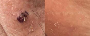
BEFORE AND AFTER SUPERFICIAL RADIATION THERAPY. RESULTS MAY VARY
Basal Cell Carcinoma
The most common form of skin cancer is basal cell carcinoma (BCC), which—as the name implies—forms in the basal cells that sit at the bottom of the outermost layer of skin. By some estimates, more than 4 million people are diagnosed with BCC in the United States each year. Often appearing as a sore, bump, or patch of discolored skin, BCC rarely grows beyond the area it first appears, making treatment fairly straightforward. This does not mean that diagnosed—or even suspected—basal cell carcinoma should be ignored, but it does mean the patient has many options, especially if the problem is caught early. While treatment options for this type of skin cancer can include topical medications or surgical excision, superficial radiation is the recommended approach, offering an effective and less invasive solution.
Squamous Cell Carcinoma
The next most common form of skin cancer, squamous cell carcinoma (SCC), develops in the uppermost layers of the skin. Doctors diagnose more than a million new cases in the United States annually, and the disease appears to be on the rise. Patients dealing with SCC will likely notice scaly, crusty, or bleeding growths, sores, or patches. These lesions most typically develop on skin that is routinely exposed to the sun, but can appear anywhere on the body. Left untreated, the cancer cells have a greater likelihood of spreading. As with BCC, squamous cell carcinoma may be managed with topical medications or surgery; however, superficial radiation is often the recommended treatment, providing an effective and less invasive option in many cases.
Melanoma
Least common of the three main types of skin cancer is melanoma, which is also the most fatal form of the disease. Melanoma develops in the cells that make pigment, which sit in the basal layer of the epidermis. Because of this, growths caused by melanoma can appear in a range of colors, but are frequently brown or black. These small tumors often resemble moles. Since melanoma can develop and spread rapidly, early detection is crucial to starting an effective treatment to destroy or remove the cancer cells and try to prevent them from metastasizing. Since this skin cancer is aggressive, treatment is typically more aggressive, too, so tactics that work on BCC or SCC may not be recommended.
What Are Skin Cancer Warning Signs?
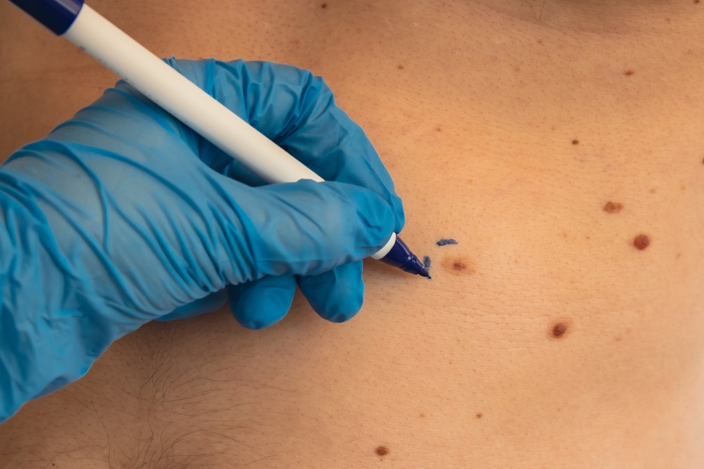
A medical doctor will make an official diagnosis of skin cancer, typically by performing a biopsy, which involves collecting a sample of skin cells from the area where skin cancer is suspected, then having them tested to confirm the presence of cancer cells. Deciding when to bring a worrisome mole, bump, or red patch in for examination is the important first step in this process.
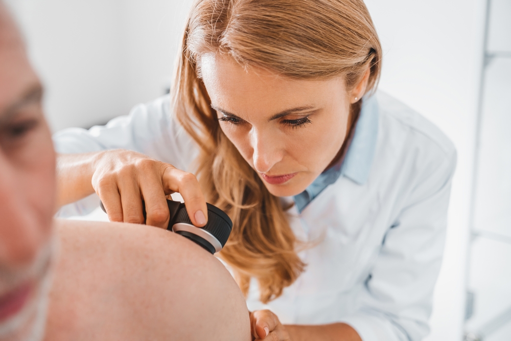
First, annual skin checks by a dermatologist can aid in the discovery of areas of concern—especially if the patient points out any particularly troublesome spots to the doctor. Patients are also always to contact a dermatologist to examine anything that may warrant a closer look. There are five factors considered to be warning signs that indicate the possible presence of skin cancer cells, which can be remembered as ABCDE:
A: Asymmetrical
The shape of a mole can be a sign of the possible presence of cancer. While many lesions are round and essentially symmetrical, the uncontrolled multiplication of cancer cells can lead to the development of asymmetrical growths. Patients who notice that one half of a mole doesn’t match the other side should ensure a dermatologist takes a closer look.
B: Border
The borders of a cancerous lesion may be ill defined or scalloped, as opposed to the clearer demarcations of a benign mole. Any spot or growth that resembles a spreading stain should be examined by a professional, who can test for skin cancer.
C: Color
Moles are typically brown, but do come in a range of colors. Typically, though, the color is uniform. A warning sign is a single mole that displays multiple shades, appearing as a mottled or mixed collection of varying browns and blacks. Red, white, and even blue may also be visible.
D: Diameter
The size of a mole matters, and though plenty of variations in diameter exist, anything larger than 6 millimeters across may be cause for concern. Smaller lesions can certainly indicate skin cancer, but any mole that reaches the size of a pencil eraser should definitely be studied.
E: Evolving
This is where self awareness and regular checks with a dermatologist come in handy. An evolving or changing mole can indicate the rapid growth of cancer cells, so a lesion that looks or feels different than it did before should trigger suspicion of skin cancer. The evolution can be in color or size. A mole may become further raised or grow in new directions. Bring an evolving mole in for an exam—especially if the evolution causes it to become asymmetrical, develop irregular borders, change colors, or grow in diameter.
Skin Cancer Treatment
As explained in the “What is Skin Cancer?” section, doctors have a range of treatments to choose from when helping a patient in the fight against skin cancer. The best potential cure depends on the patient and a host of other factors, but the main options are to kill or remove the cancer cells from the body. Dr. Ellen Turner can explain more about the specific skin cancer treatments available at her Dallas practice.
How Are Skin Cancers Treated?
Radiation for Skin Cancer
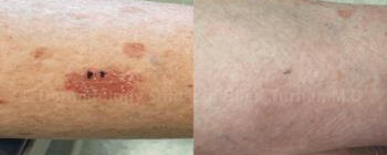
BEFORE AND AFTER SUPERFICIAL RADIATION THERAPY. RESULTS MAY VARY
A more recent and very exciting development in the treatment options for squamous cell cancer is the use of superficial radiation specifically designed to treat non-melanoma skin malignancies such as basal cell cancer and squamous cell cancer. The non-scarring, painless treatment from SRT-100 by Sensus allows for a small amount of radiation to be delivered directly into abnormal cells to include a margin in order to give the same cure rate as Mohs surgery. In particularly sensitive cosmetic areas such as the nose, ear, or eyelid, or even in areas such as the front of the lower leg, SRT-100 provides an excellent final result, minus the scarring. Many patients are opting for this therapy, and Dr. Ellen Turner’s patients are fortunate to have access to this newer technology in the Dallas/Fort Worth area.
Topical Creams for Skin Cancer
When squamous cell cancers are diagnosed at biopsy as superficial—only existing in the topmost layers of the skin—possible treatment options include topical therapy such as 5- fluorouracil, a chemotherapy cream that can be applied to the skin for a period of a few weeks. When 5-FU is used, the treated area reacts with redness, swelling, and crusting.
When basal cell cancers are biopsied and found to be superficial, they can be treated with a topical therapy such as imiquimod. Similar to 5-FU, imiquimod can induce redness, swelling, and crusting as it treats the malignant basal cells.
Surgical Excision for Skin Cancer
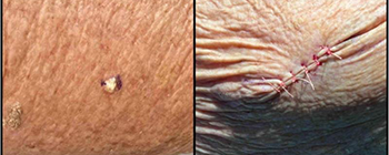
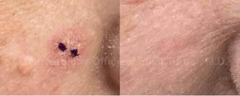
BEFORE AND AFTER SURGICAL TREATMENT FOR SKIN CANCER. RESULTS MAY VARY
This is a simple procedure performed by the dermatologist who performed the initial biopsy. Typically, biopsy-proven basal cell or squamous cell cancer on areas such as the cheek, neck, chest, arms, or back can be locally anesthetized in order to remove the skin cancer with a surrounding margin. The American Academy of Dermatology recommends the surgeon include a 5mm margin on all sides around the visualized abnormal cells so that even non-visual cells will be completely removed. Most common is the removal of an ellipse or football-shaped area so that the closure is a straight line, which will develop into a linear scar. Scars remodel themselves and continue to improve in appearance for a period of four to six months after the procedure.
Electrodessication and Curettage for Skin Cancer
This skin cancer treatment, known as ED&C, destroys skin cancer cells two ways, but only applies to basal cell carcinoma. Since squamous cell cancers may potentially involve hair follicles, and ED&C does not reach all the way down to the level of follicles, the treatment is not appropriate for squamous cell carcinoma.
ED&C involves local anesthesia to the area to be treated, plus a 4mm margin. Once the skin is numb, Dr. Ellen Turner can use a special surgical tool called a curette to remove layers of affected tissue. Then, the hyfrecator, which carries an electrical current, causes additional destruction, while stopping pinpoint bleeding. Two additional curettage passes, each followed by electrodessication using the hyfrecator, are performed, and the remaining healthy skin cells are left behind. No sutures are placed after an ED&C procedure, and the skin is allowed to heal in a process that can take up to six weeks, depending on the location of the skin cancer. Some areas are appropriate for ED&C, and others are not. Dr. Turner can advise her patients as to whether their skin cancer is amenable to treatment with electrodessication and curettage.
Mohs Surgery for Skin Cancer in Dallas
Mohs surgery requires specific criteria that a dermatologist, such as Dr. Ellen Turner, will know. She works directly with patients to determine the best treatment approach for their particular squamous cell cancer, depending on its location. Mohs surgery is often used for areas such as the nose, ear, or eyelid, where there is little excess tissue to remove. This technique involves removing an initial layer of skin, which is immediately examined for cancer cells. Additional layers are taken as needed until all cancer cells are eliminated, and closure may sometimes require a flap or graft of skin. While Dr. Turner does not offer Mohs surgery, she recommends superficial radiation as a highly effective and less invasive alternative for most patients.
Regardless of how a patient and Dr. Ellen Turner elect to treat a basal cell cancer or a squamous cell cancer, it is important to continue to monitor all skin cancer patients by performing a simple and painless full body skin exam twice yearly, or every six months in order to catch any new or changing lesions early, and get the best possible outcome.
Medical Dermatology
Please contact the dermatology office of Dr. Ellen Turner at (214) 373-7546 or online for more information on the treatment of basal cell and squamous cell skin cancers in the Dallas area.
Follow Us
Join us on social media to keep current with the latest practice news and information vital for anyone who cares about skin health and beauty.
Contact Us
Follow Us
Join us on social media to keep current with the latest practice news and information vital for anyone who cares about skin health and beauty.