

Trusted Diagnosis and Non-Invasive Treatment for the Most Common Skin Cancer in Dallas and Irving
Expert care from board-certified dermatologist Dr. Ellen Turner, with advanced treatment options including scar-free Superficial Radiation Therapy.
UNDERSTANDING BASAL CELL CARCINOMA
What Is Basal Cell Carcinoma and Why Early Diagnosis Matters
Basal cell carcinoma (BCC) is the most common form of skin cancer and the most frequently diagnosed cancer in humans. It develops in the basal cells of the epidermis, most often on sun-exposed areas such as the face, ears, neck, scalp, shoulders, and back. At the Dermatology Office of Dr. Ellen Turner in Dallas and Irving, we help patients recognize, diagnose, and treat BCC with personalized care.
The Skin Cancer Foundation estimates that 3.6 million cases of BCC are diagnosed in the United States each year, and 1 in 5 Americans will develop some form of skin cancer during their lifetime. Most BCCs are highly treatable when detected early. Although BCC rarely spreads to other parts of the body, it can slowly damage the skin and underlying tissue if left untreated.
Dr. Turner has extensive experience diagnosing and treating skin cancer, guiding patients through every step with clarity and reassurance. Whether a lesion is BCC or another condition, our priority is an accurate diagnosis followed by a treatment plan tailored to your health, lifestyle, and cosmetic goals.
BCC at a Glance
- ✓The most common form of skin cancer in the United States
- ✓Approximately 3.6 million U.S. cases diagnosed each year
- ✓Grows slowly and rarely spreads, but can cause local damage if untreated
- ✓Highly treatable when diagnosed early, with multiple effective options
1 in 5
Americans develop skin cancer
~80%
Of all skin cancers are BCC
PATIENT RESULTS
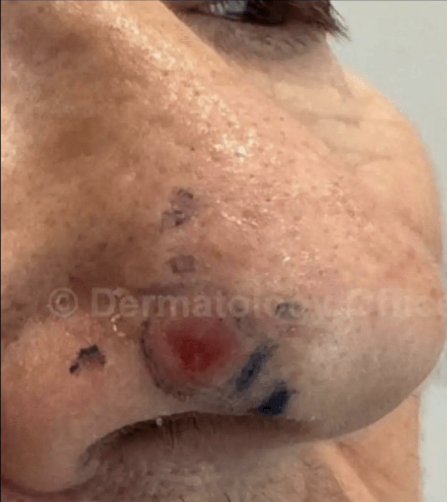
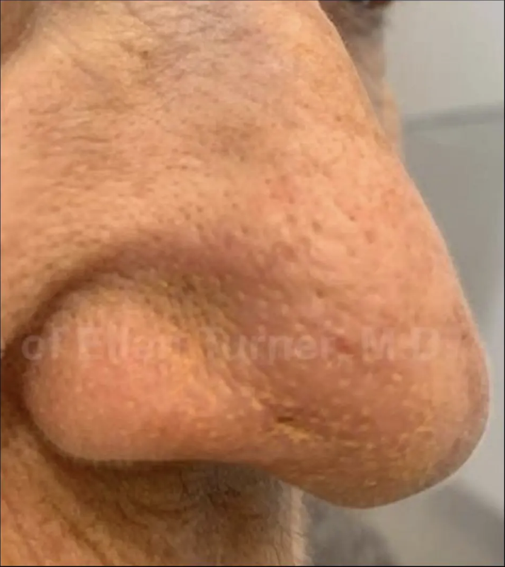
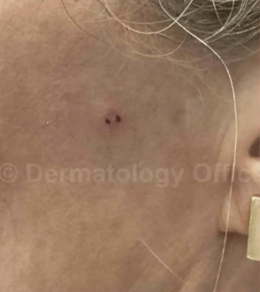
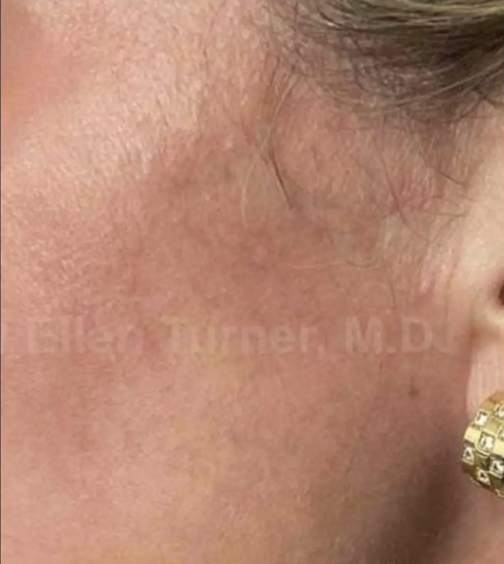
Real Results from Dr. Turner’s Dallas and Irving Patients
Actual patients treated for basal cell carcinoma with Superficial Radiation Therapy at our Dallas and Irving offices. Individual results may vary.
WHEN TO SEE A DERMATOLOGIST
How to Recognize the Warning Signs of Basal Cell Carcinoma
BCC does not always look the same, and its many disguises are one of the reasons a professional evaluation matters. If a spot on your skin looks or feels different from the rest and does not go away, do not wait — have it checked.
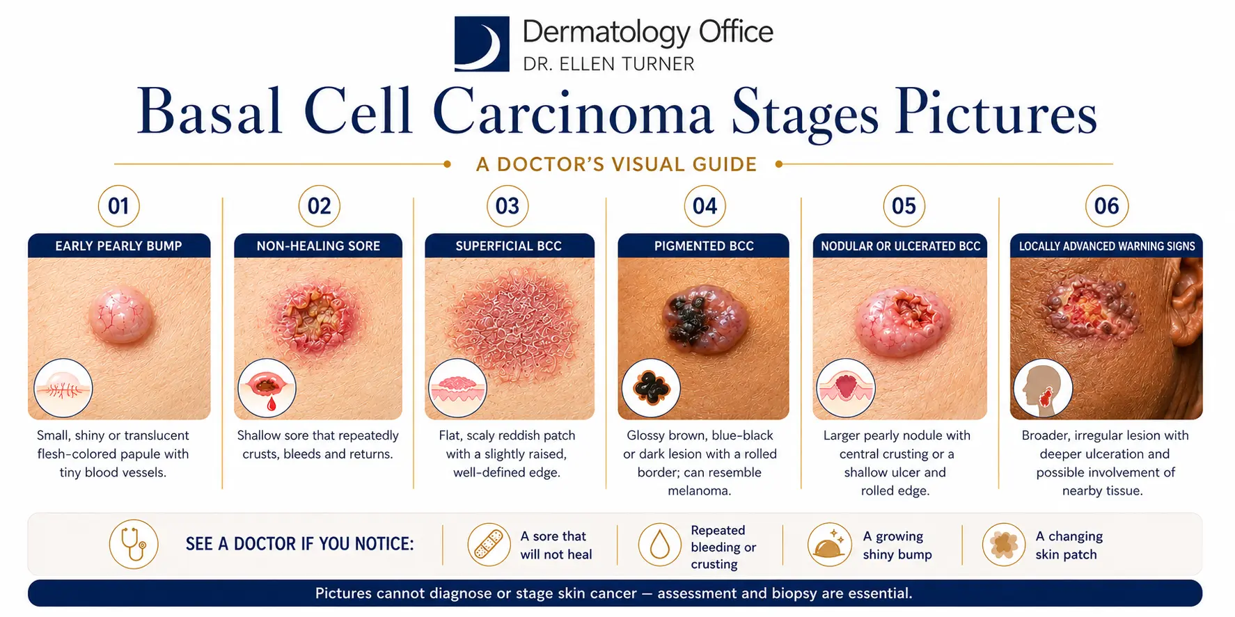
01
Pearly or Waxy Bump
Often on the face, ears, or neck. May be translucent, pink, or flesh-colored.
02
Non-Healing Sore
Bleeds, scabs, heals, then returns and lingers for weeks or months.
03
Reddish, Irritated Patch
Flat, sometimes itchy or crusty. Most often on the chest, shoulders, or back.
04
Shiny Bump or Nodule
May look like a mole or scar — skin-colored, pink, or brown in darker skin tones.
05
Rolled-Edge Growth
Slightly raised border with an indented center that may crust or bleed.
If you notice any of these signs, contact our Dallas or Irving office for an evaluation. Almost all BCCs are highly treatable when identified early.
SUBTYPES
The Many Forms of Basal Cell Carcinoma
Basal cell carcinoma is not a single disease. Dermatologists recognize several distinct subtypes, each with its own appearance, behavior, and best-fit treatment.
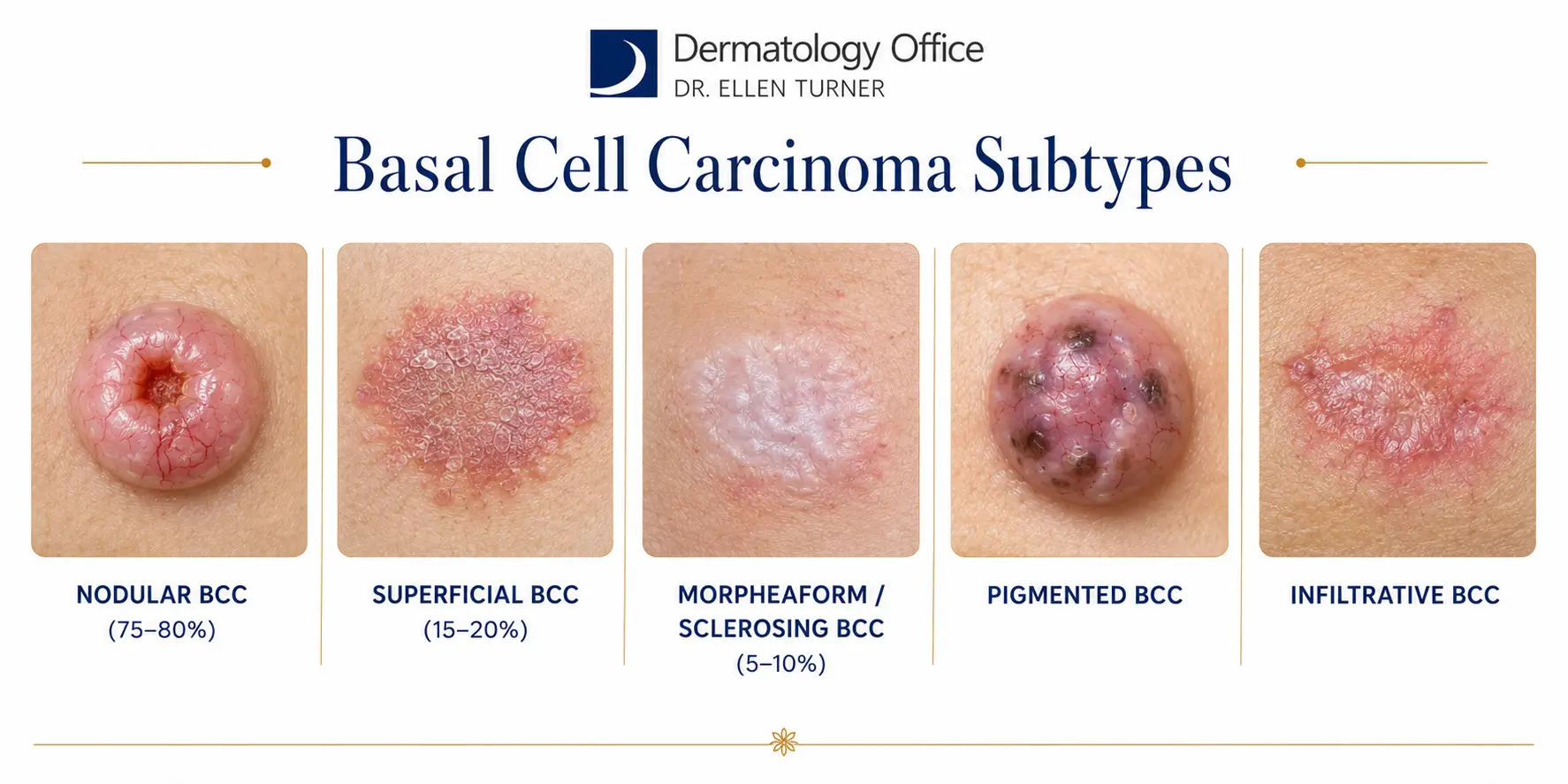
75–80% OF CASES
Nodular BCC
The most common form. A pearly, translucent bump with fine visible blood vessels crossing the surface, often with a rolled border. Most often develops on the face — especially the nose, cheeks, and ears.
15–20% OF CASES
Superficial BCC
A flat, pink or red scaly patch with a subtle raised thread-like border, most often on the trunk, shoulders, or extremities. Sometimes mistaken for eczema or psoriasis.
5–10% OF CASES
Morpheaform BCC
Also called sclerosing BCC. A white or flesh-colored scar-like patch with indistinct borders. Because it can resemble a healed scar, it is often diagnosed later than other types.
PIGMENTED VARIANT
Pigmented BCC
Similar in shape to nodular BCC but with brown, black, or blue-black pigmentation from melanin. More common in darker skin tones, and sometimes difficult to distinguish from melanoma without a biopsy.
AGGRESSIVE SUBTYPE
Infiltrative & Micronodular BCC
Aggressive subtypes with poorly defined borders that extend into surrounding tissue in thin strands or small clusters. Higher risk of recurrence and typically require more thorough treatment planning.
WHY BCC DEVELOPS
Understanding Your Risk
BCC is closely tied to lifetime ultraviolet exposure, but several other factors also raise your risk. Knowing your personal risk profile helps you and Dr. Turner plan the right screening schedule.
UV Exposure
Cumulative sun exposure and indoor tanning are the leading causes. An estimated 80.5% of BCCs are linked to UV.
Fair Skin
Individuals with light skin, light-colored eyes, and blond or red hair are at higher risk, though BCC can occur in all skin tones.
Age
Most BCCs develop after age 50, though the average age of diagnosis has been dropping in recent decades.
Personal & Family History
A prior skin cancer significantly raises the risk of another. Inherited conditions like basal cell nevus (Gorlin) syndrome also increase risk.
Immunosuppression
Organ transplant recipients and other immunosuppressed patients are at markedly higher risk of developing BCC.
Prior Radiation or Chemical Exposure
A history of ionizing radiation, arsenic exposure, or certain occupational hazards raises BCC risk.
THE DIAGNOSTIC PROCESS
How a Suspicious Lesion Becomes a Confirmed Diagnosis
A definitive BCC diagnosis always requires a biopsy. Here is what to expect at your Dallas or Irving appointment.
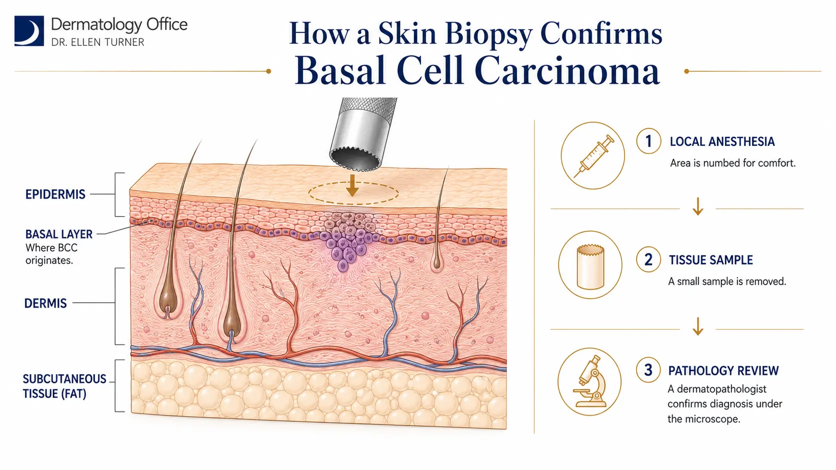
Step 1 – Clinical Skin Examination
▾
- ✓Dr. Turner performs a thorough visual examination of the lesion in question and any other suspicious areas
- ✓Your personal and family history of skin cancer, sun exposure, and any prior biopsies are reviewed
- ✓If a biopsy is recommended, the reasons are fully explained before proceeding
Step 2 – Skin Biopsy
▾
- ✓The area is cleansed and locally anesthetized so the procedure is comfortable
- ✓Depending on the lesion, a shave, punch, or excisional biopsy is performed
- ✓The tissue sample is sent to a certified dermatopathology lab for microscopic evaluation
- ✓The biopsy site is dressed and simple aftercare instructions are provided
Step 3 – Pathology Review
▾
- ✓A dermatopathologist — a physician specifically trained in skin pathology — examines the tissue
- ✓The report confirms whether cancer is present, identifies the BCC subtype, and notes depth of invasion
- ✓Superficial, nodular, and infiltrative patterns each guide treatment differently
- ✓Dr. Turner personally reviews every report before contacting the patient
Step 4 – Treatment Consultation
▾
- ✓Dr. Turner determines your customized treatment plan based on your specific diagnosis, the behavior of the basal cell carcinoma, and all available treatment options
- ✓Location, size, subtype, your health, and cosmetic priorities all factor into the plan
- ✓For most patients, several effective treatments are available, and the choice is a collaborative one
- ✓You leave with a clear next-step plan and a scheduled follow-up
TREATMENT OPTIONS
Modern, Effective Treatments for Basal Cell Carcinoma
Multiple effective treatments are available for BCC. Dr. Turner evaluates each case and, when appropriate, prioritizes non-invasive options that preserve tissue and minimize scarring while delivering excellent cure rates.
Superficial Radiation Therapy (SRT)
A precisely targeted, low-energy radiation treatment that destroys abnormal cells while preserving healthy tissue. SRT is non-invasive, painless, and scar-free, with published cure rates of 97 to 99% in clinical studies — comparable to Mohs surgery. Especially valuable for cosmetically sensitive areas such as the nose, ear, eyelid, and lower leg, and for patients who prefer or medically require a non-surgical approach.
Dr. Turner has partnered with a board-certified radiation oncologist to bring this advanced option to patients in Dallas, Irving, and the greater Dallas–Fort Worth area.
Standard Surgical Excision
Performed under local anesthesia. A 4 mm margin is removed around the tumor to ensure clearance. Effective for most BCCs, but always leaves a scar and requires wound care during healing. Reported recurrence rates around 3–4%.
Electrodesiccation & Curettage
The tumor is scraped with a curette, and an electric current is applied to destroy remaining cancer cells. The cycle is typically repeated three times. Best suited for small, superficial BCCs. Healing is slower than SRT or excision, and some scarring occurs.
Mohs Micrographic Surgery
Historically referred to as the “gold standard” for high-risk BCCs, Mohs involves removing skin layer by layer with microscopic evaluation until no cancer cells remain. Highly effective but invasive, with associated risks of bleeding, scarring, infection, and prolonged wound care. Dr. Turner reserves Mohs for cases where it is truly the most appropriate option and offers less invasive alternatives when clinically suitable.
OUR PREFERRED APPROACH
Why Superficial Radiation Therapy Is Changing How BCC Is Treated
For many patients, Superficial Radiation Therapy offers the same excellent cure rates as surgery — without the cutting, stitches, downtime, or scarring. It is one of the most exciting advancements in skin cancer care available today.

01
Non-Invasive
No incisions, no stitches, no sutures. Treatment is delivered externally with a small applicator held against the skin.
02
97–99% Cure Rates
Recent large-scale studies of image-guided SRT report freedom-from-recurrence rates exceeding 99% at 2, 4, and 6 years — comparable to Mohs surgery.
03
Scar-Free
Because SRT does not cut the skin, treated areas heal without a surgical scar — especially important on the face, ears, and lower legs.
04
Painless
Sessions are quick and painless. Most patients read or listen to music during treatment.
05
No Downtime
Patients return to their daily routine immediately after each session, with no bandages or wound care required.
06
Preserves Healthy Tissue
Radiation is precisely targeted to the tumor, sparing surrounding healthy skin — particularly valuable in cosmetically sensitive areas.
SRT is not appropriate for every patient or every lesion. Dr. Turner will discuss whether SRT fits your specific diagnosis, the tumor’s location and depth, and your medical history during your consultation.
LONG-TERM SKIN HEALTH
Prevention, Follow-Up, and Long-Term Skin Health
Successful BCC treatment is one part of the story. Preventing new skin cancers and catching any recurrence early is the other. Here is what we recommend.
01
Daily Sun Protection
Physical block SPF 30 or higher every day, reapplied every two hours in direct sun. Wide-brimmed hats, UPF clothing, and UV-blocking sunglasses add meaningful protection.
02
Avoid Indoor Tanning
Any history of indoor tanning raises BCC risk by approximately 29%, and by 69% for cases diagnosed before age 40.
03
Monthly Self-Exams
Check your skin monthly for new spots, changing lesions, or sores that will not heal. Use a mirror or ask a partner for hard-to-see areas.
04
Professional Screening
Full-body dermatology exams every 6 to 12 months for anyone with a history of skin cancer, and annually for most adults.
PATIENT QUESTIONS
Frequently Asked Questions
Is basal cell carcinoma serious?▾
How is basal cell carcinoma diagnosed?▾
Will Superficial Radiation Therapy leave a scar?▾
How long does a course of SRT take?▾
Why does Dr. Turner favor SRT over Mohs surgery?▾
Can BCC come back after treatment?▾
What are the side effects of SRT?▾
How can I prevent basal cell carcinoma?▾
Does insurance cover BCC treatment?▾
How soon should I schedule an appointment?▾
MEDICAL DERMATOLOGY | DALLAS & IRVING
Get Answers for That Suspicious Spot — Today
If you have noticed a lesion that is new, changing, non-healing, or simply looks different, do not wait. Board-certified dermatologist Dr. Ellen Turner and her team provide expert diagnosis and a full range of treatment options, including advanced Superficial Radiation Therapy, in Dallas and Irving.